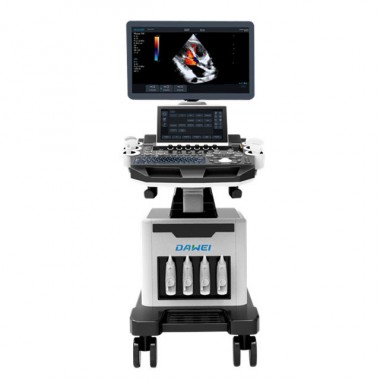
Trolley Ultrasound machine 4D echo Color Doppler scanner machine model T70
T70 ultrasound is configured with a new imaging which can significantly optimize imaging performance. It is a comprehensive imaging system engineered to meet today's most demanding needs,from deep abdominal,vascular to superficial small parts.
Adavnced ultrasonic imaging technology:
Tissue Doppler Imaging (TDI)
The Tissue doppler imaging allow you to quanittatively evaluate local myocardial movement and fuction ,with quantitative velocity parameter in TDI mode. Provide convenience for clinicans.
Auto IMT ( Intima-media thickness)
Auto measurement of anterior and posterior wall thickness providing accurate carotid status. T70 provides instant and accurate Mean and max index at the touch of a single button.
Free hand elastography
The differences in tissure responses to pressure are detected and visualized in real-time by the elastography algorithms through different graphical representations,which can be particularly helpful in analyzing breast,thyroid and musculoskeletal structures.
3D/4D imaging
T70 provides stunning fetal imaging for 2D,3D and 4D acquistions,especially for 3D/4D iaging with speed and convenience . Amazing volume performance makes T70 outshine others on volume imaging and dramatically enhances diagnostic confidence.
Panoramic imaging
The T70 provides real-time panoramic imaging for bulkly organs and masses for ease of measurement and diagnostic efficiency . Rapidly generating accurate information on the opsition of lesions.
Trapezoid imaging
Trapezoidal imaging can enhance the scanning capability for large lesions. Disconver better diagnostic information through extended view of the anatomical structure on all linear probes.
Anatomic M Mode
This advanced cardiac technology features three cursrs which can be set at any position and angle simultaneously. Reducing time needed per patient ,while maintaining diagnostic precision in addition to the standard cardiac features ,acquiring all information even in hard - to -scan situations with diffeicult heart positioning.
Auto-adaptive imaging processing
This technology used in the T70 can automatically adapt the acoustic velocity in different regions to improve the resolution and contrast.
| Parameters |
| Platforms | Windows embedded operation system( EN language) |
| Inter i5 processor |
| 4G RAM |
| Monitor | 19" medical LED monitor (1280*1024) + 10.4" touch monitor |
| Hard dish | 120G SSD+500G HDD(extendible) |
| Image models | 2D,3D,4D color/PW/CW/Power/Directional color power doppler |
| Tissue doppler,color M-model,free steering (automaticl ) |
| 3M-mode,elastrography,auto IMT,panoramic,trapezoid imaging |
| Technical Specification | Compound imaging |
| speckle reduction imaging |
| tissue harmonics imaging |
| 4D real-time |
| Automatic image optimization |
| Tissue doppler |
| Image optimization |
| Multi-beam |
| IMT |
| Trapezoidal imaging |
| i bank database |
| DICOM | Store ,print,working list,storage commitment,structured reports |
| Probe options: | Convex,linear,sector phased,Micro convex,4D volume convex |
| Applications: | Abdominal , OB/GYN,Urology,Cardiac,Vascular,Small parts |
Pediatric,MSK
|
Product Description:
Probe Options4D probe R40- Obstetrics applications
Central frequency: H5.0MHz
Multi-frequency: 3.0, H5.0, 6.0, 4.5, 3.0, 2.0MHz
Power:5-100% (arithmetic progression of 5: 5,10,15...100)
Gain:0-100
Dynamic range: 20-280% (geometric progression of 2 start from 20: 20,40,60...280)
Gray map:0-7
Frame correlation:0-4
Filtering:0-4
Image denoising:0-14
Scanning depth:3-27.3cm
Body mark:7
Scanning range:50-100% (arithmetic progression of 10 start from 50: 50,60,70...100)
Focus point: 6
Pseudo color map :0-11
Linear density:64,128,256
TSI: normal, fat, fluid, muscle
Reversal:up/down, left/right
Compound frequency: on/off
Automatic optimization: on/off
Space compound: on/off
Cardiac probe- Adult cardiac application
Frequency: 2.5, 3, 3.5, 4, H3, H4 MHz
Power:5-100% (arithmetic progression of 5: 5,10,15...100)
Gain:0-100
Dynamic range: 20-280% (geometric progression of 2 start from 20: 20,40,60...280)
Gray map:0-7
Frame correlation:0-4
Filtering:0-4
Image denoising:0-14
Scanning depth:3-27.3cm
Body mark:7
Scanning range:50-100% (arithmetic progression of 10 start from 50: 50,60,70...100)
Focus point: 5
Pseudo color map :0-11
Linear density:64,128,256
TSI: normal, fat, fluid, muscle
Reversal:up/down, left/right
Compound frequency: on/off
Automatic optimization: on/off
Space compound: on/off Image Gallery: